It’s not unusual to hear that a person invests decades of their life giving their all to support their career and family. Whether logging years in a physically demanding trade or being active in high school, collegiate, or recreational sports, a common bodily signal tends to surface in one’s forties, fifties, and beyond. The knees start to send a message. That message usually arrives as morning knee stiffness, a grinding sensation when climbing stairs, or a dull ache that settles in after a long day on their feet.
People with similar knee pain symptoms may be experiencing the early or advanced stages of knee arthritis. Arthritis is a degenerative joint condition in which the smooth cartilage that normally cushions the ends of the bones gradually wears down. Healthy cartilage allows the joint surfaces to glide smoothly during everyday movements such as walking up stairs, squatting down to pick something up, or kneeling. When that cartilage thins or becomes roughened, the bones experience increased friction and pressure where they meet. In the knee, this occurs where the femur, or thigh bone, meets the tibia, the larger bone of the shin. This deterioration can result from the natural advancement of age, the accumulated stress of a physically demanding career, overuse from years of athletic activity, or previous injuries that never fully healed.
It is worth noting that the effects of a demanding career or a vigorous athletic history do not always show up immediately. A person who ran marathons in their thirties or played collegiate sports may not feel the consequences in their knees until ten or twenty years later. The same is true for someone who spent years doing physically demanding projects around the house or going up and down stairs on a job site. Additionally, sitting for years can contribute to the deterioration of knee cartilage due to the lack of stimulus from underuse of the joint. If the lower extremities remain inactive from sitting for years over the course of a career, reduced blood flow to the supportive muscles and connective tissue in the joints can accelerate knee arthritis.
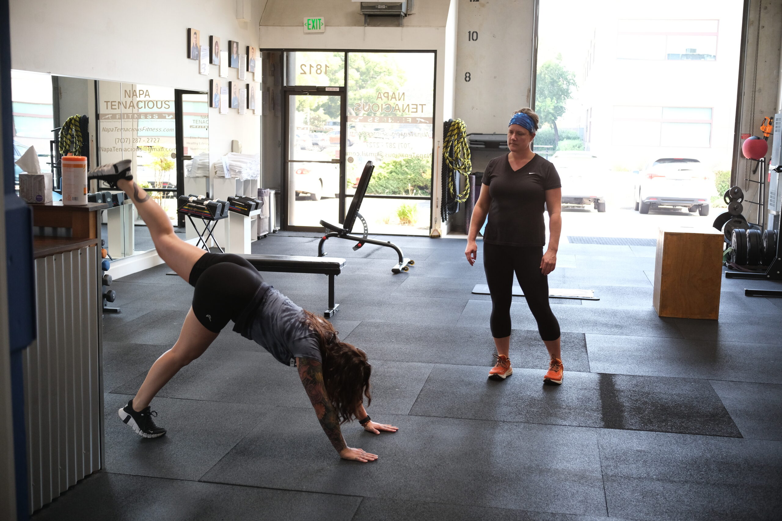
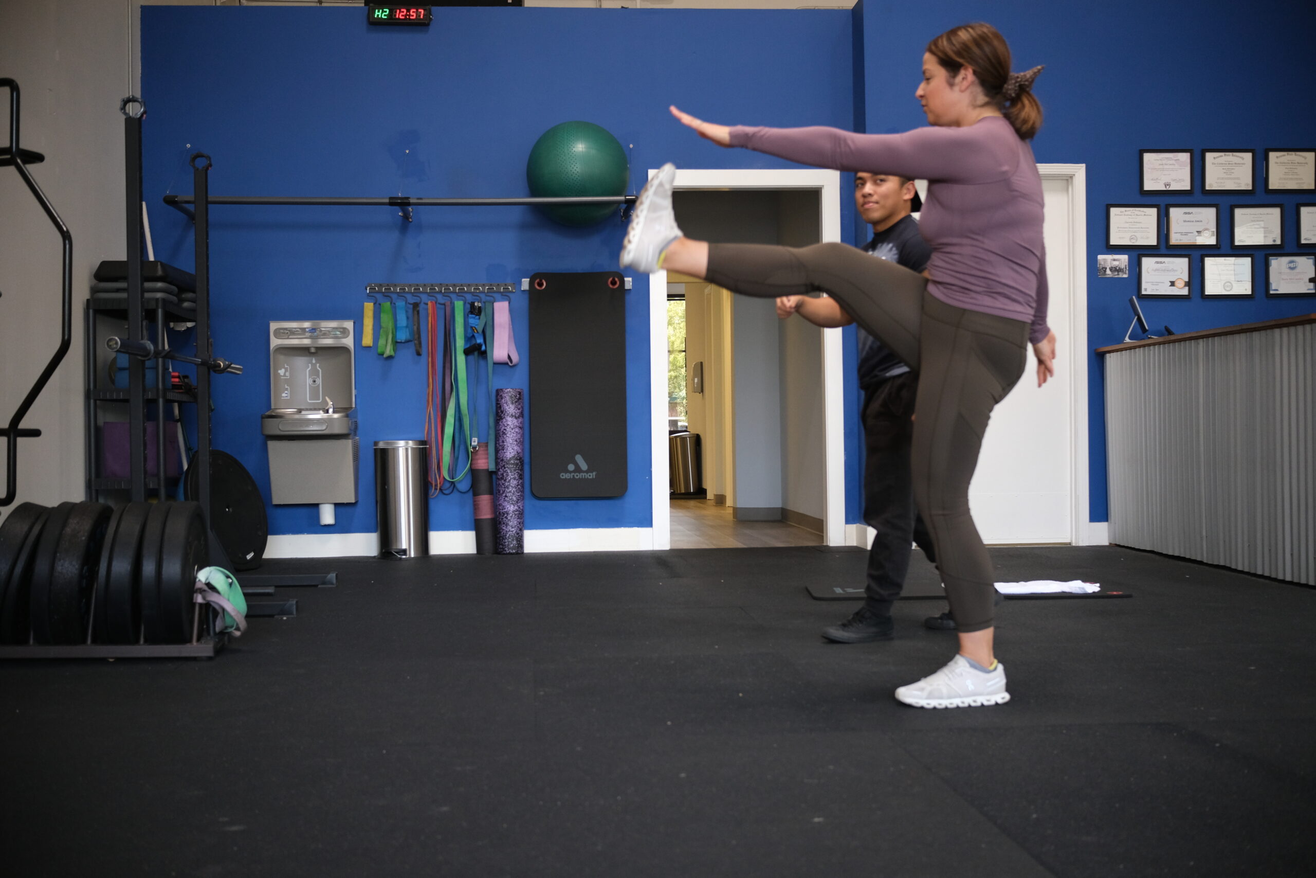
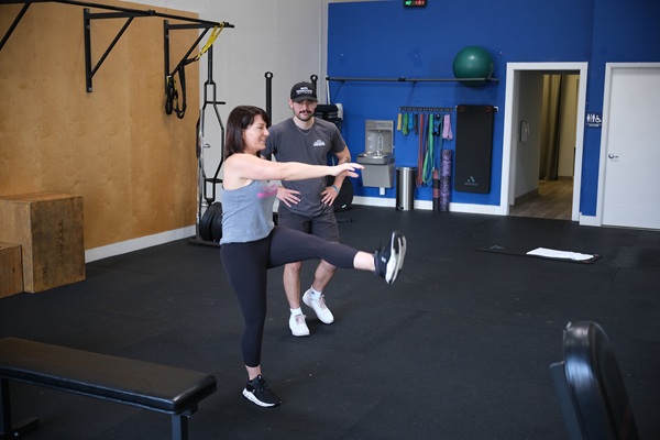
The good news is that the advancement of knee arthritis can be productively managed to offset the joint pain and dysfunction arthritis brings to the table, and it does not necessarily require cortisone shots, surgery, or expensive exercise equipment. Strengthening the muscles surrounding the knee joint is one of the most effective strategies for managing arthritis symptoms. When the muscles around the knee become stronger, they absorb a greater share of the forces generated during everyday movements such as walking, climbing stairs, or moving dynamically to get in and out of cars. This reduces the compressive load placed directly on the joint surfaces and decreases the bone-on-bone friction that causes pain.
The primary muscles that support and protect the knee joint are the quadriceps, the hamstrings, the hip adductors, and the hip abductors. The quadriceps run along the front of the thigh and are responsible for straightening the knee and keeping the kneecap tracking correctly during movement. The hamstrings run along the back of the thigh, flex the knee, and act as stabilizing anchors that prevent the joint from shifting too far forward. The hip adductors, running along the inner thigh, provide medial stability, while the hip abductors, located on the outer hip, control lateral shifting of the knee during weight-bearing activities. Together, these four muscle groups function like a set of reinforcing brackets holding the knee joint together from all sides.
One exercise we regularly prescribe for our personal training clients managing knee arthritis is the seated knee extension, which targets the quadriceps directly and assists in patellar alignment. When the quadriceps develop greater strength, endurance, and structural integrity, they help prevent the underside of the kneecap from scuffing against the surfaces of the tibia and femur, which is one of the primary sources of pain in arthritic knees. To perform the seated knee extension:
Sit on the floor with good posture. Extend one leg with the heel resting flat on the ground while keeping the other foot flat. Flex the toes of the extended leg toward the body and gently press the back of the knee toward the floor until a light muscular sensation is felt in the quadriceps. Hold briefly, then release. Repeat five to ten repetitions on each leg.
Following a weekly knee joint strengthening routine can reduce discomfort when climbing stairs, make getting up from a seated position more efficient and less painful, and noticeably reduce knee stiffness first thing in the morning and at the end of the day. When the muscles surrounding the knee grow stronger, the joint is better protected, which allows a person to be more productive and engage in the physical activities they enjoy. Developing stronger, more resilient muscles around the knee is one of the most accessible and sustainable ways to protect joint health and maintain an active, fulfilling life.
Sean McCawley, the founder and owner of Napa Tenacious Fitness in Napa, CA, welcomes questions and comments. Reach him at 707-287-2727, napatenacious@gmail.com, or visit the website napatenaciousfitness.com.